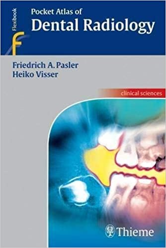
Pocket Atlas of Dental Radiology Illustrated Edition, ISBN-13: 978-3131398017
$14.99
In this age of highly specialized medical imaging, an examination of the teeth and alveolar bone is almost unthinkable without the use of radiographs. This highly informative and easy-to-read book with a collection of 798 radiographs, tables, and photos provides a myriad of problem-solving tips concerning the fundamentals of radiographic techniques, quality assurance, image processing, radiographic anatomy, and radiographic diagnosis. Information is easy to find, enabling the reader to literally get a grasp of essential new knowledge in next to no time. The dental practice team now has a pocket “consultant” at its fingertips, providing practical ways to incorporate new techniques into daily practice.
A fine-tuned didactic concept:
Each topical concept is printed compactly on a double-page spread
On the left: concise and highly instructive text
On the right: informative, high-quality illustrations
Main topics include:
- Examination strategies, radiation protection, quality assurance
- Conventional and digital radiographic techniques
- Radiographic anatomy: The problems of object localization and how to solve them
- Recent research with conventional radiography, CT, MRI, etc.
- Normal variations and pathologic conditions as viewed with the various imaging techniques
- A concise and up-to-date presentation of modern dental radiology
- Author(s): Friedrich A. Pasler, Heiko Visser
- Format: PDF
- Size: 30 MB
- 352 pages
- ISBN-13: 978-3131398017
- ISBN-10: 3131398019
- Publisher: Thieme; Illustrated edition (May 23, 2007)
- Language: English


Reviews
There are no reviews yet.